Dear All,
Thanks for your kindly comments.
Here are the summary and my responses:
1.Summary:
The high value of the R-merge
might be due to the weak diffraction, as well
as the collection method (low dose per image
and high redundancy).
The suggetions is ignore the
R-merge value, and condider the Rpim, CC1/2
and I/sigI value instead.
2. Responses to the comments:
(1) Dr. Herman Schreuder
suggested that I can use ADXV to look through
my data and see if there is some bad regions,
overload (scale only the first 30-40 frames)
or ice rings, and I also can use other
software to scale the data (xds, mosfilm
etc.)
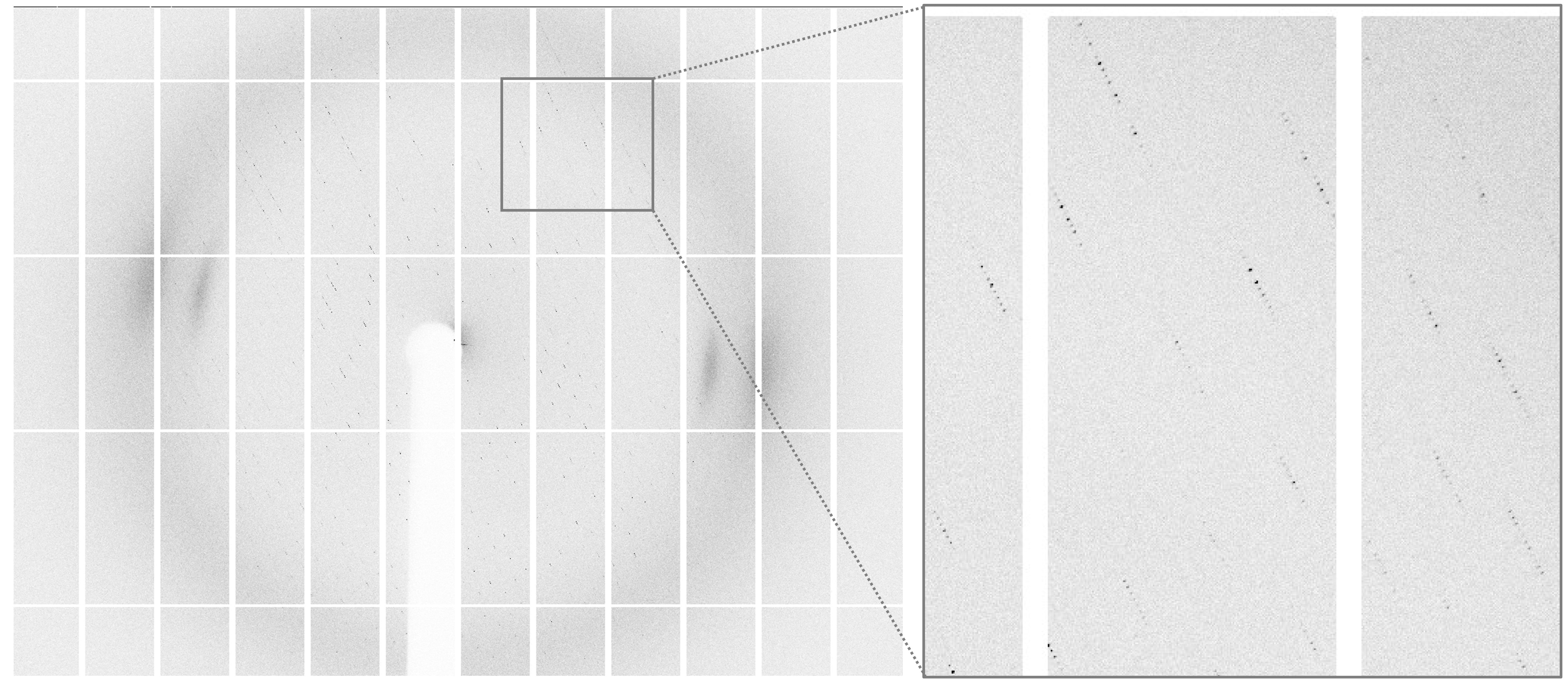
Response: Actually I have tried
these alternative ways. Indeed, the
diffraction of my crystals is pretty good (you
can see the image from attached jpg file),
there is no ice rings, no significant
radiation damage, no bad regions through the
entire frames. I have also tried to use xds to
scale it, unfortunately, the R-merge is still
high (~50%). Additionally, I also tried to
only scale part of the frames, however, the
R--merge is to ~48%.
(2) Dr. Shepard William
suggested to try mosfilm or xds, and asked the
multiplicity of the data.
Response: I have tried to scale
with XDS, but there is no improvement. The
space group is P43. I can refine the structure
to R-value 0.19, R-free 0.23, indicating that
the space group should be correct. I have also
tried to scale to P21 or P1, and there is no
improve in R-merge.
(3) Dr. Phil Evans mentioned that
Rmerge is a terrible criterion (Science, 2012,
336,1030), and CC(1/2) should be generally
considered as the best criterion. In my case,
both of the Rmerge (1.59) and CC(1/2) (0.645)
in the outer shell are acceptable. However,
the Rmerge (0.284) and CC(1/2) (0.975) in the
inner shell looks not perfect. I should
consider the radiation damage.
Response: Thanks a lot for the
comments. As you can see from the attached
figure, the diffraction is sharp, and I do not
see any significant radiation damage
(4) Dr. Ditlev Egeskov Brodersen
suggested to double check the space group and
process part of the data.
Response: as I mentioned in (1)
and (2), I have tried to only scale part of
the frames, however, the R--merge is to ~48%;
I can refine the structure to R-value 0.19,
R-free 0.23 under the current space group P43.
Moreover, scale to P21 or P1,can not improve
the R-merge significantly.
(5) Dr. Remy Loris mentioned
that a high value of R-merge indicates a wrong
symmetry or very weak data. from my data, the
reason could be the weak data as well as high
redundancy.
Response: I agree. from the
attached image, I can see the the diffraction
is sharp but weak. However, increse the
exposure time will introduce more radiation
damage....
(6) Dr. Edward A. Berry mentioned
that my data has rather high redundancy as
Rpim is much lower than Rmeas value. It could
be caused by collecting low dose per image and
making up for it with high redundancy, Dr.
Edward A. Berry suggeted to Look instead at
CC1/2 and I/sigI which seem fine.
Response: Thanks for the
comments, and I agree.
(7). Dr. Rajesh Kumar raj
suggested me to consider Rpim, CC1/2 and
I/sigI for cutting the data as Rmerge is old
approach and it is data redundancy dependent.
Thank you for your kindly help again!
Best,
Liang
I totally agree with Berry. Please consider Rpim, CC1/2 and I/sigI for cutting the data. Rmerge is old approach as it is data redundancy dependent.
Thank youRajesh
---xxxxx----With regardsRajesh K. Harijan, Ph.D.Schramm LaboratoryAlbert Einstein College of Medicine1300 Morris Park Ave., Bronx, NY 10461Tel: 718.430.2777 | Fax: 718.430.8565
The fact that chi^2 is approximately 1.0 in all shells says that the deviations are about what is expected from the error model. The fact that Rpim is much lower than Rmeas means that you have rather high redundancy. This would seem to be a case of collecting low dose per image and making up for it with high redundancy, a strategy that has been recommended to ensure a full dataset even in the case of high radiation sensitivity. In my opinion the high Rmerge is nothing to worry about. Look instead at CC1/2 and I/sigI which seem fine.
On 09/28/2018 04:09 AM, Zhang Foggy wrote:
> Dear All,
>
> Sorry for the off-topic.
>
> I recently collected a set of data. The diffraction spots are extremely sharp. However, When I used HKL3000 to scale it, I get a final resolution at 3.1A with overall R-merge ~0.54 (R-merge in the highest 3.2A-3.1A shell: 1.59). Then I solve the structure with final R value 0.19 and R free value 0.24 although I know this Rmerge value is totally unacceptable, and the density looks perfect.
>
> I also tried to collect other four set of data with different crystals. unfortunately, all of them have same problem.
>
> I ask one of my friend who is an expert in HKL3000, but he had no idea about it. Does anyone has suggestions?
>
> Here is the scale information for your review:
> Space group: P43 (I also tried P1, the Rmerge value is still similar)
>
> Shell Lower Upper Average Average Norm. Linear Square
> limit Angstrom I error stat. Chi**2 R-fac R-fac Rmeas Rpim CC1/2 CC*
> 50.00 6.67 11.6 0.9 0.3 1.165 0.191 0.284 0.198 0.052 0.975 0.994
> 6.67 5.30 4.5 0.5 0.3 0.952 0.317 0.313 0.329 0.086 0.971 0.993
> 5.30 4.63 7.3 0.7 0.5 0.961 0.293 0.297 0.304 0.081 0.975 0.994
> 4.63 4.21 7.0 0.8 0.6 0.986 0.369 0.358 0.382 0.101 0.969 0.992
> 4.21 3.91 5.6 0.8 0.6 1.040 0.522 0.491 0.541 0.142 0.955 0.988
> 3.91 3.68 4.6 0.9 0.7 1.064 0.718 0.669 0.746 0.203 0.929 0.981
> 3.68 3.49 3.5 0.9 0.8 1.092 1.059 0.986 1.101 0.299 0.882 0.968
> 3.49 3.34 2.6 0.9 0.8 1.092 1.382 1.298 1.438 0.395 0.829 0.952
> 3.34 3.21 2.1 0.9 0.8 1.084 1.543 1.489 1.614 0.468 0.772 0.933
> 3.21 3.10 1.6 0.9 0.8 1.070 1.591 1.669 1.680 0.529 0.645 0.885
> All reflections 5.0 0.8 0.6 1.048 0.538 0.487 0.559 0.153
>
> Thank you.
>
> Liang
>
>
> ------------------------------------------------------------------------------------------------------------------------------------------------------------------------------------------------------------------------------------------------------------------------------------------------------------------------------------------------------------------------------------------------------------------------------------------------------------------------------------------------------------------------------------------------------------------------------------------------------------------------------------------------------------------------------------------------------------------------------------------------------------------------------------------------------------------------------------------------------------------------------------------------------------------------------------------------------------------------------------------------------------------------------
>
> To unsubscribe from the CCP4BB list, click the following link:
> https://www.jiscmail.ac.uk/cgi-bin/webadmin?SUBED1=CCP4BB&A=1
>
########################################################################
To unsubscribe from the CCP4BB list, click the following link:
https://www.jiscmail.ac.uk/cgi-bin/webadmin?SUBED1=CCP4BB&A=1
To unsubscribe from the CCP4BB list, click the following link:
https://www.jiscmail.ac.uk/cgi-bin/webadmin?SUBED1=CCP4BB&A=1
To unsubscribe from the CCP4BB list, click the
following link:
https://www.jiscmail.ac.uk/cgi-bin/webadmin?SUBED1=CCP4BB&A=1