 |
Clinical query:
Clinico-Radiological correlation in atypical Pneumonia
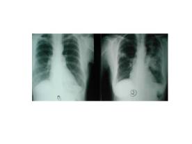
A 60-year-old lady presented to us with fever, cough and shortness of breath for 2 days. A chest X-ray done on presentation was normal (fig 1). However her symptoms persisted and a chest x-ray repeated after 2 days showed bilateral upper zone infiltrates with air broncho-grams suggestive of consolidation (fig1).
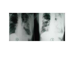
She was put on empirical antibiotics, IV penicillin, amino glycosides and macrolides. After 3 days she improved symptomatically with the fever, cough and shortness of breath decreasing but a repeat chest x-ray showed a progression of the infiltrates. (Fig2)
1) Is atypical pneumonia commonly known to present with a poor clinico radiological correlation?
2) Is there a known reason for such a phenomenon?