Dear SPMers:
I am analyzing the fMRI data using SPM5, but I am confusing with the step of normalize. When I follow the steps listed in the SPM5 manual(realign, coregister between T1 and fMRI images, segmentation of T1 image, normalization of fMRI images using the parameter file got from the segmentation step, specify and estimate), something wrong with the result in rendering the SPM.mat on the T1 anatomic image(see the attached file). However, the SPM.mat could be overlaid on the colin.img very well. Why? Can you give me any instruction about how to normalize the fMRI data to his T1 anatomic image?
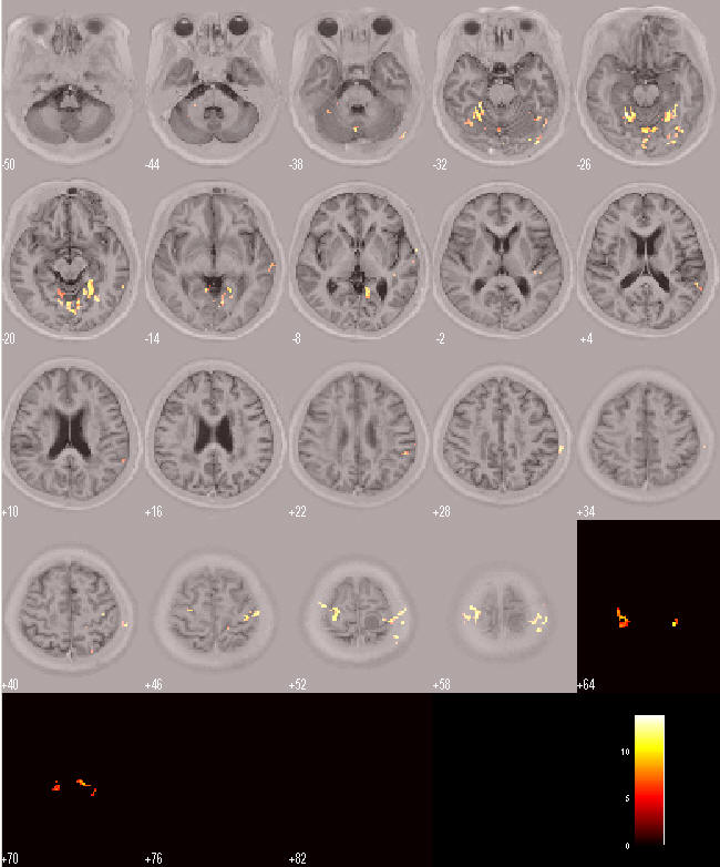
Note to the attached files:The file hands_fMRI.jpg is the picture that the activation map overlaid on the coregistered T1 image. You can see some activation is located outside the brain. The file Hands_fMRI_with_canonical is the picture that the activation map overlaid on the colin.img.
Yours sincerely,
Zhi Wang
[log in to unmask]
[log in to unmask]
No.1 Dahua Road Dongdan
The Dept. of Radiology
Beijing Hospital
Beijing,China 100730
2007-03-02
|


