Dear all,
This is a question about nearest neighbour interpolation
used in the process of normalization.
I normalized a functional dataset with this interpolation
method. The normalized images look fine, but in several
voxels, there are strange signal behaviors.
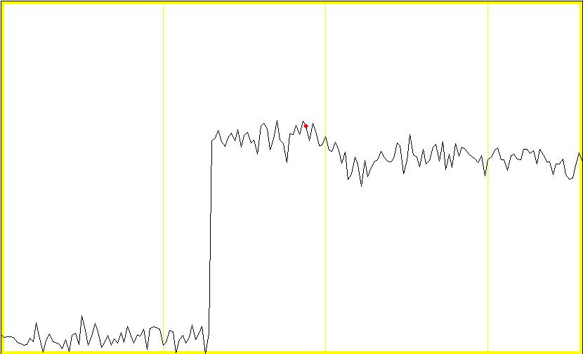
The attached jpeg image shows an example. That is a plot of
a timecourse of one voxel in the grey matter. The signal
proceeds with small ups and downs for several time points,
but around the first one-third of the timecourse, it
suddenly jumps up, and after that it proceeds normally
again.
Such an odd signal behavior is seen in many other voxels.
Some show jump-ups, some show drop-downs, and others show
both. Those voxels seem to be distributed around the whole
brain randomly.
I tried normalizing the same data set with the other two
interpolation methods, sinc and bilinear. In these two cases
no strange signal behaviors were observed.
The details of the normalization parameters were as follows:
Determin parameters & write normalized
# of subjects: 1
Image to determine parameters from: coregistered
anatomical T1 image
Images to write normalized: functional EPI
images(180scans)
Template: T1.img
# of nonlinear basis functions: 8*8*8
# of iterations: 16
Medium regularization
Interpolation: nearest neighbour
voxel sizes: 2*2*2mm
(original EPI voxel sizes: 3.75*3.75*4mm)
Any comment/suggestion would be greatly appreciated.
Kota KATANODA
Dept.Cogn.Neurosci.
Fac.Med. Univ.Tokyo
|


